dermatome and myotome pdf

Dermatomes and myotomes are key anatomical concepts representing sensory and motor nerve distributions. Dermatomes are skin areas supplied by specific spinal nerves, while myotomes are muscle groups innervated by these nerves. Understanding their organization and clinical relevance is essential for neurological examinations and diagnosing nerve root lesions. Detailed PDF guides provide comprehensive charts and maps, serving as invaluable resources for anatomy students and healthcare professionals.
1.1 Definition of Dermatomes
A dermatome is a specific area of skin supplied by sensory nerve fibers originating from a single spinal nerve root. Each dermatome corresponds to a particular spinal nerve, providing sensation to a distinct region of the body. Dermatomes are essential for understanding sensory innervation and are often visualized through detailed maps. These maps illustrate the distribution of dermatomes across the body, from the cervical (C1-C8) to the sacral (S1-S5) regions. Dermatomes are crucial in clinical settings for diagnosing nerve root lesions, as symptoms like numbness or tingling can be correlated with specific dermatomal patterns. This concept is fundamental in neurology and anatomy, aiding in precise neurological examinations and patient assessments.
1.2 Definition of Myotomes
A myotome is a group of muscles innervated by motor nerve fibers originating from a single spinal nerve root. Each myotome corresponds to specific spinal nerves, controlling voluntary movements of associated muscles. Myotomes are crucial for understanding motor innervation and are often studied alongside dermatomes. They develop from somites during embryological development, forming the foundation of muscle organization. Myotomes are categorized into proximal and distal groups, reflecting their anatomical distribution. In clinical settings, myotomes are essential for diagnosing nerve root lesions, as muscle weakness or paralysis can be linked to specific myotomal patterns. This concept is vital in neurology and physical therapy, aiding in the assessment and rehabilitation of motor functions.
1.3 Importance of Dermatomes and Myotomes in Anatomy
Dermatomes and myotomes are fundamental concepts in anatomy, providing a detailed understanding of sensory and motor nerve distributions. Dermatomes map skin areas supplied by specific spinal nerves, while myotomes identify muscle groups innervated by these nerves. This knowledge is crucial for correlating symptoms with nerve root involvement, aiding in the diagnosis of conditions like numbness, tingling, or muscle weakness. In clinical practice, dermatomes and myotomes guide neurological examinations and physical therapy interventions. They also serve as essential tools for educating anatomy students and healthcare professionals, offering a clear framework for understanding nerve function and its relationship to the body’s structure. Their precise mapping enhances diagnostic accuracy and treatment planning in neurology and rehabilitation medicine;
Embryological Origin
Dermatomes and myotomes originate from somites, paired structures in the embryo that develop into skin, muscles, and connective tissues. Somites form segmentally, influencing nerve connections and innervation patterns.
2.1 Development of Dermatomes from Somites
Dermatomes develop from somites, paired structures in the embryo that form along the head-to-tail axis. Each somite differentiates into three main components: the dermatome, myotome, and sclerotome; The dermatome contributes to the skin, while the myotome forms muscles, and the sclerotome develops into bones and cartilage. During development, somites segment and migrate, establishing the pattern for sensory innervation. Dermatomes are innervated by spinal nerves, which maintain their segmental connections. This organization is crucial for understanding nerve root function and diagnosing lesions. The migration of dermatome cells ensures proper alignment with spinal nerves, forming the basis for dermatomal maps used in clinical practice. This developmental process is detailed in dermatome and myotome PDF guides, which provide visual and textual insights for anatomy students and professionals.
2.2 Development of Myotomes from Somites
Myotomes originate from somites, paired embryonic structures that segment along the body axis. Each somite differentiates into a dermatome, myotome, and sclerotome. The myotome gives rise to skeletal muscles, which are innervated by spinal nerves. During development, somites migrate and establish segmental nerve connections, ensuring motor innervation follows muscle migration. This process is crucial for forming the organized pattern of muscle groups supplied by specific nerve roots. The development of myotomes is closely linked to dermatomes, as both arise from the same somitic precursors; Understanding this embryological origin is vital for correlating muscle function with nerve root activity, as detailed in dermatome and myotome PDF guides, which provide comprehensive insights for anatomy and clinical applications.
2.3 Role of Somites in Human Embryology
Somites are paired, segmental structures in the early embryo that play a pivotal role in human development. They form along the head-to-tail axis and differentiate into three main components: dermatomes, myotomes, and sclerotomes. Dermatomes contribute to the skin, myotomes to skeletal muscles, and sclerotomes to vertebrae and ribs. Somites also give rise to tendons and cartilage, essential for musculoskeletal structure. Their segmental organization establishes the body’s metameric pattern, influencing nerve distribution and muscle formation. This embryological process is foundational for understanding dermatomes and myotomes, as detailed in dermatome and myotome PDF guides, which highlight their clinical and anatomical significance in nerve root function and muscle innervation.

Dermatomes

Dermatomes are areas of skin supplied by specific spinal nerve roots, providing sensory innervation. They follow a segmental pattern, crucial for diagnosing nerve root lesions. Detailed PDF guides offer comprehensive charts and maps, aiding in understanding their distribution and clinical relevance for neurological assessments and education.
3.1 Anatomy of Dermatomes
Dermatomes are discrete areas of skin innervated by specific spinal nerve roots, forming a map of sensory distribution across the body. Each dermatome corresponds to a particular nerve root, with their arrangement following a segmental pattern. Dermatomes are crucial for understanding sensory innervation and diagnosing nerve root lesions. They are organized into cervical, thoracic, lumbar, and sacral regions, with distinct boundaries that overlap minimally. The dermatomes of the upper and lower limbs form loops, such as C5-T1 and L4-S1, while the trunk and head have more linear distributions. Detailed PDF guides provide visual representations of these dermatomes, aiding in clinical and educational applications. This anatomical organization is vital for correlating sensory deficits with specific nerve roots.
3.2 Distribution of Dermatomes Across the Body
Dermatomes are organized segmentally, covering the entire body from the cervical region to the sacral area. Each dermatome corresponds to a specific spinal nerve root, forming a continuous map of sensory innervation. The cervical dermatomes (C1-C8) cover the neck, shoulder, and upper limbs, while thoracic dermatomes (T1-T12) span the torso. Lumbar (L1-L5) and sacral (S1-S5) dermatomes innervate the lower limbs and pelvic region. This distribution follows a predictable pattern, with dermatomes forming loops around the limbs (e.g., C5-T1 and L4-S1). While the general arrangement is consistent, minor variations exist between individuals. Detailed PDF guides provide visual representations of this distribution, aiding in clinical diagnosis and educational understanding of sensory innervation patterns.
3.3 Dermatomes of the Upper Limbs
The upper limbs are innervated by dermatomes corresponding to cervical and thoracic spinal nerve roots. Dermatomes from C5 to T1 cover the arm, forearm, and hand, forming a distinct pattern. C5 dermatome covers the lateral arm, while C6 extends to the lateral forearm and thumb; C7 dermatome innervates the middle finger and posterior arm, and C8 covers the medial forearm and little finger. T1 dermatome supplies the medial arm and axilla. This segmental distribution is crucial for clinical assessments, as it helps identify nerve root lesions. Detailed PDF guides provide clear maps of these dermatomes, aiding in precise diagnosis and understanding of sensory innervation in the upper limbs.
3.4 Dermatomes of the Lower Limbs
The lower limbs are primarily innervated by dermatomes corresponding to lumbar and sacral spinal nerve roots. The dermatomes from L1 to S5 cover distinct areas of the legs and feet. L1 dermatome covers the groin and medial thigh, while L2 extends to the anterior thigh. L3 dermatome innervates the knee and lower thigh, and L4 covers the shin and medial ankle. L5 dermatome supplies the dorsum of the foot, and S1 dermatome covers the posterior thigh and lateral foot. S2 to S5 dermatomes innervate the perianal region. These dermatomes are essential for clinical assessments, aiding in the diagnosis of nerve root lesions. Detailed PDF guides provide clear maps of these dermatomes, facilitating accurate neurological evaluations and understanding of sensory distribution in the lower limbs.
3.5 Dermatomes of the Trunk and Head
The trunk and head are innervated by dermatomes corresponding to cervical and thoracic spinal nerve roots. Cervical dermatomes (C1-C8) cover the neck, shoulder, and upper back, while thoracic dermatomes (T1-T12) innervate the chest, abdomen, and back. The dermatomes of the head are unique, as they are supplied by branches of the trigeminal nerve (V1, V2, V3). These dermatomes are crucial for assessing sensory function in neurological examinations. Detailed PDF guides provide maps of these dermatomes, aiding in the correlation of symptoms with specific nerve root involvement. Understanding these dermatomes is essential for diagnosing conditions affecting the trunk and head, such as nerve root lesions or spinal injuries;

Myotomes
Myotomes are groups of muscles innervated by specific spinal nerve roots, essential for motor function. They develop from somites and are crucial for understanding motor innervation patterns in anatomy and clinical assessments.
4.1 Anatomy of Myotomes
Myotomes are groups of muscles innervated by specific spinal nerve roots, playing a crucial role in motor function. Each myotome corresponds to a particular nerve root and is essential for understanding motor innervation patterns. Derived from somites during embryological development, myotomes follow a segmental organization, reflecting their origin. This segmentation allows for precise motor control and is vital for clinical assessments of muscle weakness. The anatomy of myotomes is closely linked to their developmental origins, as they migrate and merge to form adult muscles. Their structure and function are fundamental to diagnosing nerve root lesions and understanding motor deficits in neurological examinations.
4.2 Distribution of Myotomes Across the Body
Myotomes are distributed segmentally across the body, corresponding to specific spinal nerve roots. Each myotome represents a group of muscles innervated by a single nerve root, following a proximal-to-distal pattern. In the upper limbs, myotomes are primarily associated with cervical and thoracic nerve roots (C5-T1), while lower limb myotomes correspond to lumbar and sacral roots (L2-S2). The trunk’s myotomes are organized between thoracic (T2-T12) and lumbar (L1) levels. This distribution aligns with embryological development, where somites give rise to segmental muscle groups. Understanding this organization is crucial for clinical assessments, as it helps correlate muscle weakness with specific nerve root lesions, aiding in precise diagnoses and targeted therapies.
4.3 Myotomes of the Upper Limbs
Upper limb myotomes are groups of muscles innervated by specific cervical and thoracic nerve roots (C5-T1). Each myotome corresponds to a particular spinal nerve, controlling distinct movements. For example, the C5 myotome governs shoulder abduction, while C6 controls elbow flexion. The distribution follows a proximal-to-distal pattern, with C5-T1 myotomes contributing to movements like shoulder rotation, elbow extension, and wrist flexion. This segmental organization is vital for clinical assessments, as muscle weakness in specific areas can indicate nerve root lesions; Detailed PDF guides provide charts mapping these myotomes, aiding in precise diagnoses and targeted rehabilitation strategies for upper limb injuries or conditions.
4.4 Myotomes of the Lower Limbs
Lower limb myotomes are muscle groups innervated by lumbar and sacral nerve roots (L2-S2). Each root corresponds to specific movements, such as hip flexion (L2-L3), knee extension (L3-L4), and ankle dorsiflexion (L4-S1). These myotomes follow a proximal-to-distal distribution, enabling precise motor control. For example, the L5 myotome controls ankle dorsiflexion and toe extension, while S1 governs ankle plantarflexion. Clinical assessments of lower limb myotomes help identify nerve root lesions, guiding targeted rehabilitation. Detailed PDF guides provide charts mapping these myotomes, aiding in accurate diagnoses and treatment plans for conditions like sciatica or spinal injuries. Understanding these patterns is crucial for anatomy students and healthcare professionals.
4.5 Myotomes of the Trunk and Head
The trunk and head myotomes are primarily innervated by cervical nerve roots (C1-C4). These myotomes control essential movements such as neck flexion, extension, and rotation. The C1-C2 myotomes regulate subtle head movements, while C3-C4 influence larger neck muscles. Additionally, the diaphragm, innervated by the phrenic nerve (C3-C5), is crucial for respiration. Damage to these myotomes can result in respiratory weakness or limited cervical mobility. Clinical assessments of these areas are vital for diagnosing nerve root lesions. Detailed PDF guides provide charts mapping these myotomes, aiding in accurate diagnoses and treatment plans for conditions like cervical spondylosis. Understanding these patterns is essential for anatomy students and healthcare professionals, ensuring precise clinical correlations and interventions.
Clinical Relevance
Dermatomes and myotomes are crucial in diagnosing nerve root lesions and muscle weaknesses. They guide physical therapy and neurological examinations, correlating symptoms with specific spinal nerve involvement, aiding precise treatment plans.
5.1 Dermatomes and Myotomes in Neurological Examinations
Dermatomes and myotomes play a vital role in neurological examinations, enabling clinicians to assess sensory and motor function. By testing specific dermatomes, healthcare providers can identify nerve root lesions, while myotome evaluations reveal muscle weakness. These assessments are crucial for diagnosing conditions like spinal cord injuries or nerve impingements. Detailed PDF guides provide clear maps and charts, aiding in precise testing and interpretation. Regular use of these tools ensures accurate correlations between symptoms and nerve involvement, guiding effective treatment plans and physical therapy interventions. This systematic approach enhances diagnostic accuracy and patient outcomes in clinical practice.
5.2 Correlation of Dermatomes and Myotomes with Spinal Nerve Roots
Dermatomes and myotomes are closely linked to specific spinal nerve roots, providing a clear anatomical map for clinical correlation. Each dermatome corresponds to a particular spinal nerve, enabling precise identification of sensory deficits. Similarly, myotomes represent muscle groups innervated by individual nerve roots, aiding in the detection of motor weaknesses. This correlation is essential for diagnosing nerve root lesions, as symptoms often align with specific dermatomal or myotomal patterns. Detailed PDF guides illustrate these relationships, facilitating accurate assessments. Understanding this connection is crucial for localizing neurological deficits and guiding targeted interventions, making it a cornerstone of clinical neurology and physical therapy practices.
5.3 Clinical Tests for Assessing Dermatomes
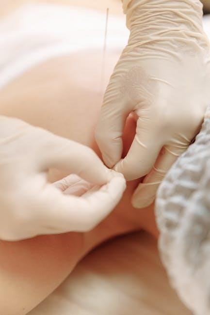
Clinical tests for assessing dermatomes involve evaluating sensory function across specific skin areas. Common methods include the pinprick test to assess pain perception and light touch assessment using a soft brush or cotton swab. These tests help identify deficits corresponding to specific dermatomes. For example, diminished sensation in the C6 dermatome suggests a potential issue with the sixth cervical nerve root. Detailed PDF guides provide standardized techniques and reference points for accurate testing. These assessments are critical in neurological examinations to localize nerve root lesions and guide targeted interventions. By systematically testing dermatomes, clinicians can map sensory abnormalities, aiding in precise diagnoses and effective treatment plans.
5.4 Clinical Tests for Assessing Myotomes
Clinical tests for assessing myotomes focus on evaluating motor function and muscle strength. Key assessments include manual muscle testing, where strength is graded on a scale from 0 to 5. Reflex testing, such as knee-jerk and ankle reflexes, provides insights into myotome integrity. Observing for muscle atrophy or fasciculations can indicate chronic nerve root compromise. Gait analysis and range-of-motion exercises also help identify weaknesses linked to specific myotomes. These tests are essential for diagnosing nerve root lesions and guiding rehabilitation. Detailed PDF guides offer standardized protocols for myotome evaluation, ensuring accurate and reproducible assessments. By systematically testing myotomes, clinicians can pinpoint motor deficits and develop targeted treatment plans.
Dermatome and Myotome Mapping
Dermatome and myotome mapping involves detailed charts correlating skin areas and muscle groups with specific nerve roots. These tools aid in diagnosing nerve root lesions and are essential for anatomy education and clinical practice.

6.1 Dermatome Maps
Dermatome maps are visual tools that illustrate the specific areas of skin innervated by individual spinal nerve roots. These maps are crucial for understanding the sensory distribution of dermatomes across the body. They are often used in clinical settings to correlate symptoms with nerve root involvement, aiding in the diagnosis of conditions like nerve root lesions or spinal injuries. Detailed dermatome maps are available in PDF guides, which include charts and diagrams for each spinal level, from C1 to S5. These resources are particularly valuable for anatomy students, neurologists, and physical therapists. By referencing these maps, professionals can accurately assess sensory deficits and plan appropriate interventions. They are also widely used in educational materials to help learners memorize dermatome patterns.
6.2 Myotome Charts
Myotome charts are detailed visual representations of muscle groups innervated by specific spinal nerve roots. These charts are essential for understanding the motor distribution of myotomes across the body. They are widely used in clinical and educational settings to identify patterns of muscle weakness associated with nerve root lesions. Myotome charts are often included in dermatome and myotome PDF guides, providing a clear and organized reference for professionals and students. By mapping the motor functions of each spinal nerve, these charts aid in the assessment of motor deficits and the planning of rehabilitation strategies. They complement dermatome maps, offering a comprehensive understanding of both sensory and motor nerve root functions.
6.3 Combined Dermatome and Myotome Maps
Combined dermatome and myotome maps provide a comprehensive visual representation of both sensory and motor nerve root distributions. These maps integrate dermatomes, which depict skin areas supplied by specific spinal nerves, with myotomes, which illustrate muscle groups innervated by the same nerves. This integration allows for a holistic understanding of nerve root function and its clinical implications. Such maps are particularly useful in diagnosing nerve root lesions, as they correlate sensory deficits with motor weaknesses. They are often included in dermatome and myotome PDF guides, serving as invaluable tools for healthcare professionals and anatomy students. By visualizing both dermatomes and myotomes together, these maps enhance the ability to assess and interpret neurological symptoms effectively.
Practical Applications

Dermatomes and myotomes are essential for diagnosing nerve root lesions and assessing muscle weakness. They guide physical therapy interventions and serve as educational tools for anatomy students and professionals.
7.1 Use of Dermatomes in Diagnosing Nerve Root Lesions
Dermatomes are crucial in diagnosing nerve root lesions by correlating sensory deficits with specific spinal nerve roots. For instance, numbness in the C6 dermatome suggests a C6 nerve root lesion. Clinical tests, such as pinprick and light touch assessments, are used to evaluate sensory loss within dermatomal patterns. This helps pinpoint the affected nerve root, guiding further investigations like MRI or EMG. Dermatome maps, often provided in PDF guides, offer clear visual references for clinicians to accurately localize lesions. These tools are indispensable in neurology and physical medicine, enabling precise diagnosis and targeted treatment plans for patients with suspected nerve root pathology.
7.2 Use of Myotomes in Diagnosing Muscle Weakness
Myotomes play a vital role in diagnosing muscle weakness by linking specific muscle groups to their corresponding spinal nerve roots. For example, weakness in elbow flexion indicates a C5 or C6 myotome involvement. Clinical assessments, such as manual muscle testing, are used to evaluate strength within myotomal patterns. This helps identify nerve root lesions or injuries affecting motor function. Myotome charts, often included in dermatome and myotome PDF guides, provide detailed mappings of muscle groups and their innervating nerves. These resources are essential for clinicians to accurately diagnose and manage conditions like radiculopathy or spinal injuries, ensuring targeted rehabilitation strategies for patients with motor deficits.
7.3 Role of Dermatomes and Myotomes in Physical Therapy
Dermatomes and myotomes are essential tools in physical therapy for designing targeted rehabilitation programs. By understanding the sensory and motor distributions, therapists can identify areas of impairment and develop exercises to restore function. For instance, identifying specific dermatomal patterns helps address sensory deficits, while myotome analysis guides motor retraining. This knowledge enables therapists to create personalized treatment plans, improving outcomes for patients with nerve injuries or spinal conditions. Additionally, dermatome and myotome maps from PDF guides aid in tracking progress and refining therapies. This approach ensures a focused and effective rehabilitation process, enhancing recovery and functional mobility for patients with neurological or musculoskeletal impairments.
Educational Resources
Comprehensive dermatome and myotome PDF guides offer detailed charts and maps for anatomy students and professionals. Online tools and recommended reading materials provide interactive learning and in-depth insights.
8.1 Dermatome and Myotome PDF Guides
Dermatome and myotome PDF guides provide detailed, organized charts and maps, enhancing both clinical and educational applications. These resources are ideal for anatomy students and professionals, offering clear insights into the sensory and motor distributions of spinal nerve roots. The guides cover dermatomes for each spinal nerve from C1 to S5, along with corresponding myotomes, making them indispensable for correlating symptoms with nerve root involvement. They include assessment techniques for evaluating nerve root injuries in the upper and lower extremities, as well as reflexes and special tests. Easy to access and comprehensive, these PDFs are essential for understanding dermatomes and myotomes, serving as valuable tools for medical education and practice.
8.2 Online Tools for Learning Dermatomes and Myotomes

Online tools and resources provide interactive and accessible ways to learn dermatomes and myotomes. Websites like Geeky Medics offer comprehensive guides, including dermatome maps and myotome charts, to aid in understanding nerve root distributions. Kenhub and The Noted Anatomist provide detailed tutorials, videos, and downloadable PDFs for anatomy students. These platforms often include quizzes, flashcards, and interactive diagrams to enhance learning. Additionally, virtual patients and OSCE stations simulate real-world clinical scenarios, helping learners apply dermatome and myotome knowledge in practical settings. These tools are invaluable for medical students, professionals, and educators seeking to master dermatomes and myotomes effectively.
8.3 Recommended Reading for Anatomy Students
For anatomy students, recommended reading includes comprehensive guides and resources that detail dermatomes and myotomes. Websites like Geeky Medics and Kenhub offer detailed tutorials, videos, and downloadable PDFs. These resources provide clear charts, maps, and explanations, making complex concepts accessible. Additionally, The Noted Anatomist offers premium tutoring memberships and free online resources, including OSCE stations and flashcards. These materials are tailored for both students and professionals, offering practical insights into dermatomes and myotomes. They are essential for mastering nerve root anatomy and its clinical applications, ensuring a strong foundation for neurological examinations and diagnoses.
Dermatomes and myotomes are fundamental concepts in anatomy, essential for understanding nerve distribution and clinical diagnostics. Their detailed mapping in PDF guides provides invaluable resources for students and professionals alike.
9.1 Summary of Key Concepts

Dermatomes and myotomes are anatomical regions supplied by specific spinal nerve roots. Dermatomes represent areas of skin receiving sensory innervation, while myotomes are groups of muscles receiving motor innervation. Both originate from somites during embryological development. Understanding their distribution and function is crucial for diagnosing nerve root lesions and muscle weaknesses. Dermatomes follow a segmental pattern, forming loops around limbs, while myotomes exhibit proximal-to-distal organization. Clinical tests, such as sensory assessments and muscle strength evaluations, rely on these concepts. PDF guides provide detailed maps and charts, aiding in education and professional practice. These resources are indispensable for correlating symptoms with nerve root involvement, making them essential tools in anatomy and clinical medicine.
9.2 Future Directions in Dermatome and Myotome Research
Future research on dermatomes and myotomes may focus on refining their clinical applications, particularly in diagnosing nerve root lesions and muscle weaknesses. Advances in imaging and neurophysiological testing could enhance the precision of dermatome and myotome mapping. Additionally, integrating these concepts into artificial intelligence systems for automated diagnosis could revolutionize clinical practice. Educational tools, such as interactive 3D models and virtual reality, may improve learning outcomes for anatomy students. Furthermore, exploring the embryological origins of dermatomes and myotomes in greater detail could provide insights into developmental anomalies. These advancements will likely deepen our understanding and improve the practical application of dermatomes and myotomes in medicine and education.